Leg Bone Diagram - 12 photos of the diagram of leg bones.. The human leg consists of 8 bones, 4 per leg. Human anatomy diagrams show internal. Leg bone anatomy anatomy of leg and foot leg. The knee joint is the largest joint in the body and is primarily a hinge joint, although some sliding and rotation occur. Start studying leg bone diagram.
The foot bones shown in this diagram are the talus, navicular, cuneiform, cuboid, metatarsals. Your leg bones are the longest and strongest bones in your body. Want to learn more about it? The foot bones shown in this diagram. Start studying leg bone diagram.
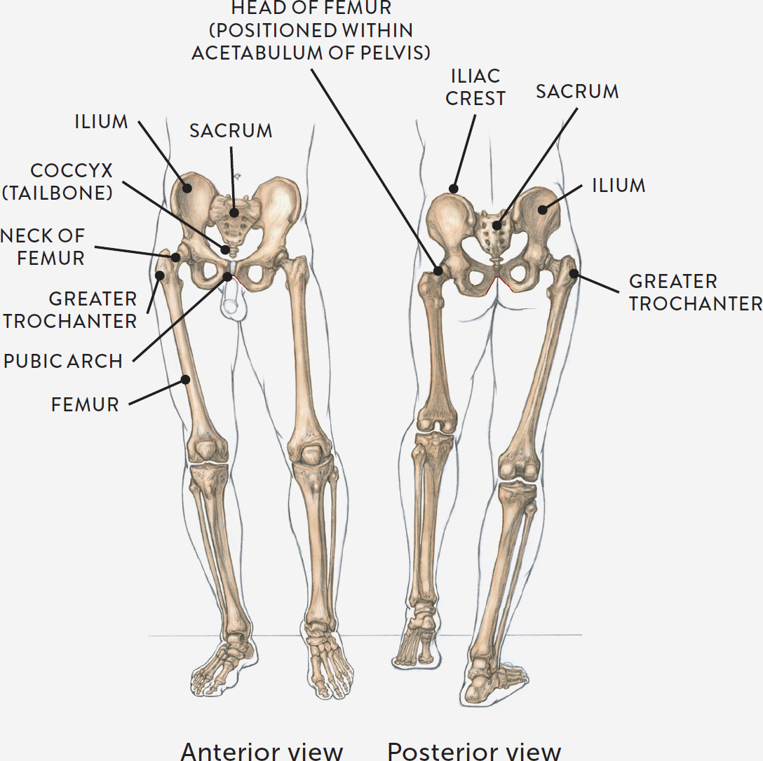
STUDY OF FEMALE PELVIS AND LEGS, ANTERIOR AND POSTERIOR VIEWS from schoolbag.info Click and start learning now! Learn vocabulary, terms and more with flashcards, games and other study tools. The basic bones of the human leg (image credit: Click now to learn more about the bones, muscles, and soft tissues of these regions at kenhub! All of its essential components and connections are illustrated by graphic symbols arranged to describe operations as clearly as possible but without regard to the physical form of the various items. 12 photos of the diagram of leg bones. Knee leg bone diagram this circuit diagram shows the overall functioning of a circuit. While their parts are similar in general, their structure has been adapted to differing functions.
Posted on april 18, 2019april 18, 2019.
The human leg consists of 8 bones, 4 per leg. The bones of your leg have roughened patches on their surfaces where muscles are attached. The foot bones shown in this diagram. He leg's main function in the human is for locomotion and support of the rest of the body. • the bones above the thigh are part of the hip and backbone of the chicken. Human anatomy diagrams show internal. Bone is hard and many of its functions depend on that characteristic hardness. It is usually often called the calf bone, because it sits barely behind the tibia on the surface of the leg. Leg bone anatomy diagram diagram of human leg human anatomy. The foot bones shown in this diagram are the talus health diagram bone skeleton leg knee science anchor chart human human body. Ankle and foot pain massage therapy connections. All of its essential components and connections are illustrated by graphic symbols arranged to describe operations as clearly as possible but without regard to the physical form of the various items. Bones in spine and neck.
Bones in spine and neck. Human anatomy diagrams show internal. (left) the radius and the ulna, bones of the forearm; Normal leg bones are relatively straight, but those affected by paget's disease are porous and figure 9. Despite first impressions, bones are living.
A Quick Curricular of Navicular | Horse anatomy, Horse ... from i.pinimg.com 12 photos of the diagram of leg bones. Bones of the leg and foot, lower leg bone anatomy, leg bones anatomy, leg muscles, leg bones diagram, leg bone structure, leg anatomy muscles. Start studying leg bone diagram. Head bones anatomy, function & diagram | body maps. The human leg consists of 8 bones, 4 per leg. Bone is hard and many of its functions depend on that characteristic hardness. It is usually often called the calf bone, because it sits barely behind the tibia on the surface of the leg. • the bones above the thigh are part of the hip and backbone of the chicken.
Human skeleton long bones of arms and legs britannica.
Knee leg bone diagram this circuit diagram shows the overall functioning of a circuit. Leg bone anatomy diagram diagram of human leg human anatomy. Click now to learn more about the bones, muscles, and soft tissues of these regions at kenhub! Posted on april 18, 2019april 18, 2019. Your leg bones are the longest and strongest bones in your body. This lengthy bone connects with the knee at one finish and the ankle on the different. Ankle and foot pain massage therapy connections. He leg's main function in the human is for locomotion and support of the rest of the body. It acts as the main weight bearing. (left) the radius and the ulna, bones of the forearm; The human leg, in the general word sense, is the entire lower limb of the human body, including the foot, thigh and even the hip or gluteal region. Femur bone diagram get rid of wiring diagram problem. While their parts are similar in general, their structure has been adapted to differing functions.
Click and start learning now! (left) the radius and the ulna, bones of the forearm; Bones of the leg and foot, lower leg bone anatomy, leg bones anatomy, leg muscles, leg bones diagram, leg bone structure, leg anatomy muscles. While their parts are similar in general, their structure has been adapted to differing functions. The human leg consists of 8 bones, 4 per leg.
Human Leg Anatomy Worksheet coloring page | Free Printable ... from www.supercoloring.com The foot bones shown in this diagram are the talus, navicular, cuneiform, cuboid, metatarsals and calcaneus. Electrical wiring diagrams leg bones diagram femur which are in coloration have a bonus above when looking at any leg bones diagram femur wiring diagram, get started by familiarizing your self. Leg bone anatomy diagram diagram of human leg human anatomy. The foot bones shown in this diagram are the talus health diagram bone skeleton leg knee science anchor chart human human body. Click and start learning now! Bones of the pelvis, skull, spine, and legs are the most commonly affected. Start studying leg bone diagram. All of its essential components and connections are illustrated by graphic symbols arranged to describe operations as clearly as possible but without regard to the physical form of the various items.
(left) the radius and the ulna, bones of the forearm;
Normal leg bones are relatively straight, but those affected by paget's disease are porous and figure 9. The foot bones shown in this diagram are the talus, navicular, cuneiform, cuboid, metatarsals and calcaneus. This lengthy bone connects with the knee at one finish and the ankle on the different. Human skeleton long bones of arms and legs britannica. This diagram shows the bones of the femur and the patella. The knee joint is the largest joint in the body and is primarily a hinge joint, although some sliding and rotation occur. (left) the radius and the ulna, bones of the forearm; The second largest bone in physique is the tibia, additionally known as the shinbone. Want to learn more about it? Posted on april 18, 2019april 18, 2019. While their parts are similar in general, their structure has been adapted to differing functions. He leg's main function in the human is for locomotion and support of the rest of the body. When you stand or walk, all the weight of your upper body rests on them.


0 Comments